Cerebral Blood Flow
Cerebral Blood Flow Heading link

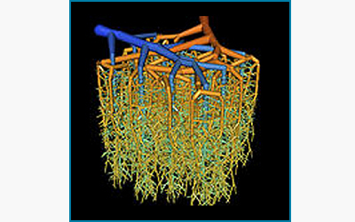
Hemodynamics and Oxygen Exchange in the Mouse Cerebral Microcirculation
Ian Gould, Grant Hartung, and Andreas Linninger
A detailed map of the mouse cerebral cortex with all individual neurons and glial cells as well as the embedded microcirculatory blood flow network was constructed In collaboration with Dr. Kleinfeld at UCSD. Our lab created a morphologically accurate, physiologically consistent, multi-scale computer model of the entire cerebral microcirculation to predict blood flow, hematocrit distribution, red blood cell saturation, neuronal and glial oxygen tension including mitochondrial oxygen turnover. The cellular simulations elucidates the functional role of the angioarchitecture in supplying oxygen to brain cells after neuronal firing. Computational models at the cellular level are expected to improve our understanding of changes in hemodynamics and oxygen extraction occurring in cerebrovascular diseases such as micro infarcts, vascular dementia, cerebral vasospasm, epilepsy and Alzheimer’s.
Computational Study of Changes to the Human Microcirculation in the Diseased and Aging Brain Heading link
Computational Study of Changes to the Human Microcirculation in the Diseased and Aging Brain
Ian Gould and Andreas Linninger
Our lab developed a novel biphasic blood flow model to predict blood flow and resulting oxygenation in the human secondary cortex. We synthesized on the computer virtual representations of the human cortex including all capillaries, as well as individual brain cells. Computer predictions of oxygen exchange and solute transfer across the blood brain barrier help elucidate brain function of the neurovascular unit and explain brain diseases as well as the aging brain.
Whole Brain Flow Quantification Heading link
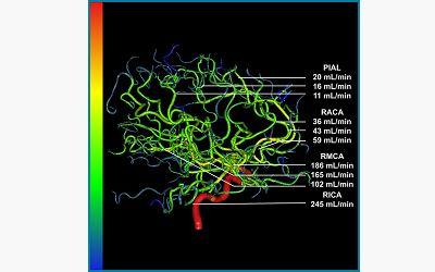
Whole Brain Flow Quantification
Chih-Yang Hsu and Andreas Linninger
Digital subtraction angiography (DSA) can currently only be used for qualitative assessment of blood distribution patterns. We are developing new imaging software to infer volumetric blood flow rates from dynamic DSA signals. The novel method enables the creation of patient-specific maps of cerebral blood flow in every blood vessel visible by angiography. Volumetric blood flow estimation is feasible from large arteries in the Circle of Willis to pial arteries and cortical veins.